On the way of scientific research and technical development, MinFound Medical accomplished PET detector digitalization. By now MinFound Medical's PET/CT solutions include middle-range and high-end solutions with comprehensive clinical applications, including widely required function of radiotherapy positioning mode. Let’s break down main characteristics in details.

MinFound PET detector digital technology utilizes digital signal processing hardware as a means to achieve accurate sampling of scintillation pulses. Compared with traditional analog detector technology, digital detector technology has better time, energy and spatial resolution, can obtain higher quality clinical images, and improve lesion conspicuousness, lesion clarity and diagnostic confidence. At the same time, the application of digital detector technology can shorten the acquisition time or reduce the dose of radioactive tracers [1][2][3][4], thereby improving the acquisition efficiency or reducing the radiation dose.

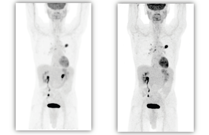
Clinical image based on a device's Clinical image based on MinFound ScintCare
PMT-PET analog detector PET/CT 730T SiPM digital detector

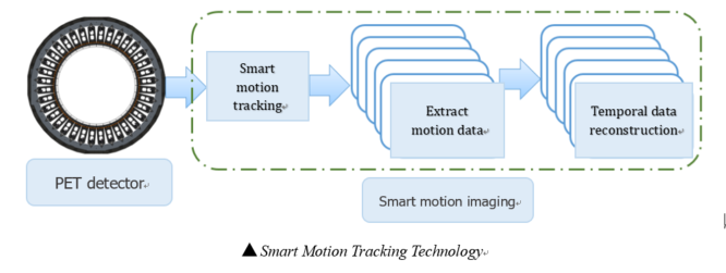
MinFound Medical’s ScintCare PET/CT series are adhered to independent innovation. The intelligent motion artifact correction technology uses the statistical difference of the moving ray source in the projection domain to extract motion information and intelligent tracking to realize the correction of head, breathing, heart and other motion artifacts. Intelligent motion artifact correction technology can eliminate image quality degradation or motion artifacts caused by patient motion without adding any clinical burden (extra scan), thus providing more accurate and realistic images for clinic[5 ][6].
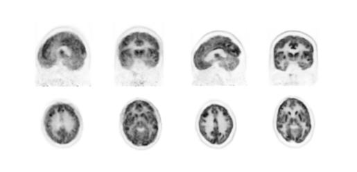
▲Images before and after intelligent head motion artifact correction
(Left image: front, right image: rear)
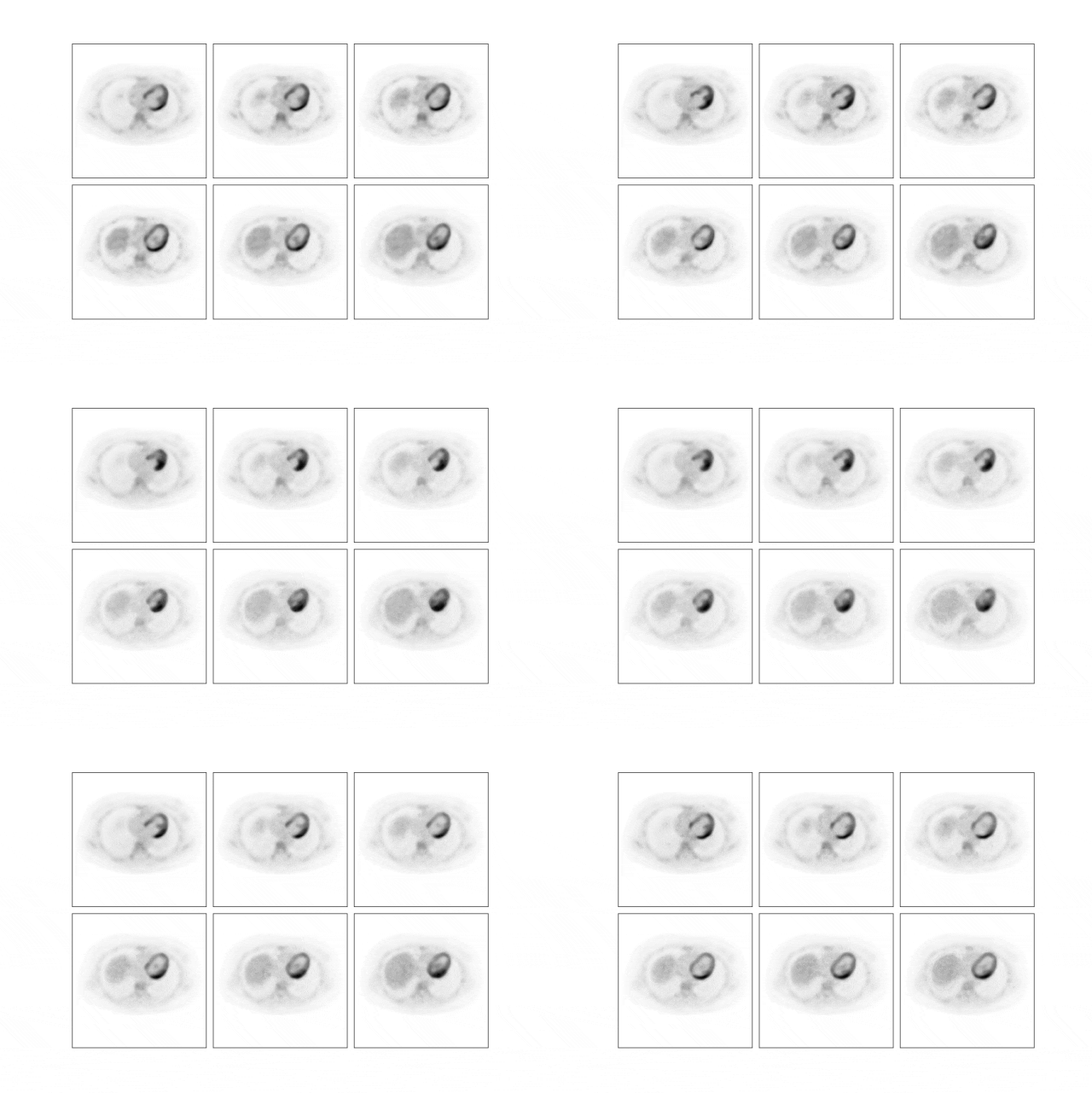
▲Smart Heart Motion Tracking Image

The radiation brought by PET/CT scanning often causes concerns regarding "nuclear discoloration". The standard dose of traditional PET/CT imaging is generally 0.12~0.15mCi/kg. If the dose or scanning time is reduced, the image quality and focus of PET images will be affected. Accuracy of detection and quantification.
The "DosePower" low-dose module developed by MinFound Medical employed in its PET/CT products adopts a hybrid iterative intelligent reconstruction method based on the data domain and the image domain, which can well preserve the details and contrast of the image,furthermore, effectively solve the over-smoothing problems caused by general algorithms. It singificantly facilitates to lower clinical injection dose (0.04~0.07mCi/kg), faster scanning (<60s), and more accurate images[7][8][9][10][11].
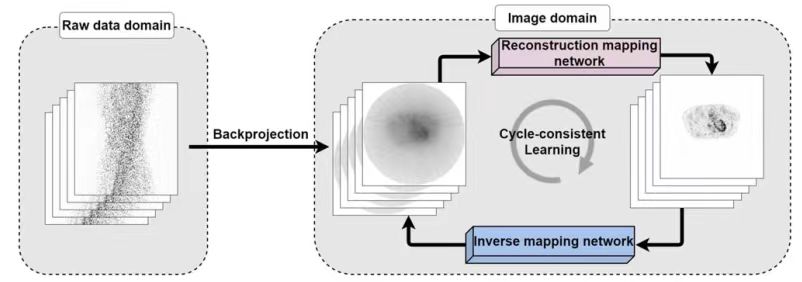
▲Schematic diagram of intelligent low-dose reconstruction algorithm
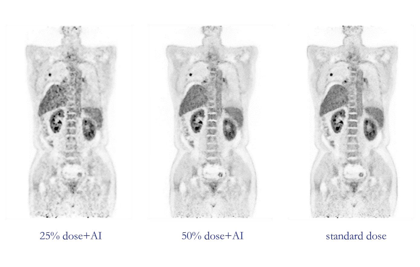
▲Comparison of Clinical Image Effects

The image quality of PET/CT equipment depends on the performance status of key components, so regular quality control of PET/CT is required to ensure the best condition of the equipment. Traditional quality control methods use external radiation sources and require on-site operation by quality control personnel, which greatly increases the workload and radiation dose of on-site quality control personnel. MinFound Medical’s PET/CT intelligent passive quality control and calibration technology is based on LYSO background radioactivity to realize PET quality control data and calibration data reservation-style intelligent collection and analysis, which solves the problem of traditional quality control relying on external radiation sources and manual intervention. Optimize customer experience and ensure equipment status in real time [12][13][14];
 MinFound Medical's PET/CT intelligent full-time calibration technology is based on real-time detector data collection and modeling during the clinical scanning process, and realizes real-time monitoring and intelligent calibration of PET detector performance such as time, energy and temperature, ensuring the authenticity and accuracy of data in the clinical process. Effectiveness, thereby improving the image signal-to-noise ratio [15] [16].
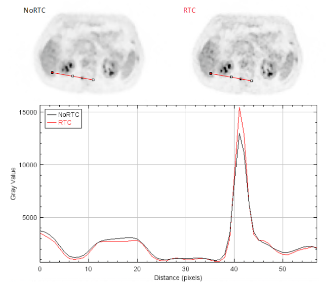
▲Comparison of clinical images before and after intelligent full-time calibration
DISCLAIMER: THE ACTUAL IMAGES QUALITY MIGHT EXCEL THE PROVIDED CLINICAL IMAGES.
[1] Schillaci, O., Urbano, N. Digital PET/CT: a new intriguing chance for clinical nuclear medicine and personalized molecular imaging. Eur J Nucl Med Mol Imaging 46, 1222–1225 (2019).
[2] Deng Z, Deng Y, Chen G. Design and Evaluation of LYSO/SiPM LIGHTENING PET Detector with DTI Sampling Method. Sensors (Basel). 2020 Oct 15;20(20):5820.
[3]Thomas K. Lewellen. The Challenge of Detector Designs for PET. Nuclear Medicine and Molecular Imaging. August 2010
[4] Pietro P.Calò a, Fabio Ciciriello a, Savino Petrignani a. SiPM readout electronics, Nuclear Instruments and Methods in Physics Research, Section A: Accelerators, Spectrometers, Detectors and Associated Equipment. May 2019(57-58).
[5] Koivumaki T, Nekolla SG, Furst S, Loher S, Vauhkonen M, Schwaiger M, et al. An integrated bioimpedance—ECG gating technique for respiratory and cardiac motion compensation in cardiac PET. Phys Med Biol. 2014;59(21):6373–85
[6] Sarah J McQuaid, Tryphon Lambrou and Brian F Hutton. A novel method for incorporating respiratory-matched attenuation correction in the motion correction of cardiac PET–CT studies. Phys Med Biol.2011;56:2903-2915
[7] Bao Yang, Long Zhou, Ling Chen, Lijun Lu, Huafeng Liu and Wentao Zhu. Cycle-consistent learning-based hybrid iterative reconstruction for whole-body PET imaging. Phys. Med. Biol. 67 (2022) 085016.
[8]Kui Zhao, Size Gao, Yaofa Wang , Hongwei Ye. Study of low-dose PET image recovery using supervised learning with CycleGAN. PLoS One 2020 Sep 4; 15(9).
[9] Yang Q, Yan P, Zhang Y, Yu H, Shi Y, Mou X, et al. Low-dose CT image denoising using a generative adversarial network with Wasserstein distance and perceptual loss. IEEE transactions on medical imaging. 2018; 37(6):1348–1357
[10] Kaplan S, Zhu YM. Full-Dose PET Image Estimation from Low-Dose PET Image Using Deep Learning:a Pilot Study. Journal of digital imaging. 2018; p. 1–6
[11] Chen KT, Gong E, de Carvalho Macruz FB, Xu J, Boumis A, Khalighi M, et al. Ultra–Low-Dose 18F-Florbetaben Amyloid PET Imaging Using Deep Learning with Multi-Contrast MRI Inputs. Radiology. 2018;290(3):649–656
[12] Alva-Sánchez, H., Zepeda-Barrios, A., Díaz-Martínez, V.D.et al. Understanding the intrinsic radioactivity energy spectrum from 176Lu in LYSO/LSO scintillation crystals. Sci Rep 8, 17310 (2018)
[13] Enríquez-Mier-y-Terán, F.E., Ortega-Galindo, A.S., Murrieta-Rodríguez, T.et al. Coincidence energy spectra due to the intrinsic radioactivity of LYSO scintillation crystals. EJNMMI Phys 7, 21 (2020).
[14] Conti M,Eriksson L, Rothfuss H, et al. Characterization of 176Lu background in LSO-based PET scanners. Phys Med Biol. 2017; 62: 3700– 3711.
[15] Arabi, H., Zaidi, H. Non-local mean denoising using multiple PET reconstructions.Ann Nucl Med 35, 176–186 (2021)
[16] Liu, Y., Gao, Mj., Zhou, J.et al. Changes of [18F]FDG-PET/CT quantitative parameters in tumor lesions by the Bayesian penalized-likelihood PET reconstruction algorithm and its influencing factors. BMC Med Imaging 21, 133 (2021). https://doi.org/10.1186/s12880-021-00664-7